Anatomy Of The Upper Chest Area : oitf-chest-extrinsic | Integrative Works / According to frederic delavier, author of the strength training anatomy books, with bilateral work, both shoulders are driven backward supporting the weight.
Anatomy Of The Upper Chest Area : oitf-chest-extrinsic | Integrative Works / According to frederic delavier, author of the strength training anatomy books, with bilateral work, both shoulders are driven backward supporting the weight.. The upper posterior border of the heart is formed by the left atrium. Atlas of anatomy of the human body: The chest is part of a larger group of pushing muscles found in hemi diaphragm normal chest anatomy lateral chest xray colon gas trachea oblique fissure horizontal fissure rt. Anatomy of peritoneum and mesentery. I will therefore split the chest up into three parts:
Diagram of ganglionic areas numbered 1 to 14, used in clinical practice in. According to frederic delavier, author of the strength training anatomy books, with bilateral work, both shoulders are driven backward supporting the weight. It describes the theatre of events. We're looking at the anatomy of an upper endoscopy. Enlargement will result in bulging of the.
Neck And Chest Anatomy - Anatomy Drawing Diagram from thoracickey.com It is a rare but serious condition, with the potential to cause vascular compromise of the upper limb. Seen clearly crossing the upper part of each lung field. Anatomy is to physiology as geography is to history: Related posts of anatomy of the chest area. It describes the theatre of events. Upper back pain and chest pain can occur together. For the purpose of description the lungs are divided into zones: Radiological anatomy of the chest please view our editing file before studying this lecture to the black parts resemble the trachea.
Related posts of anatomy of the chest area.
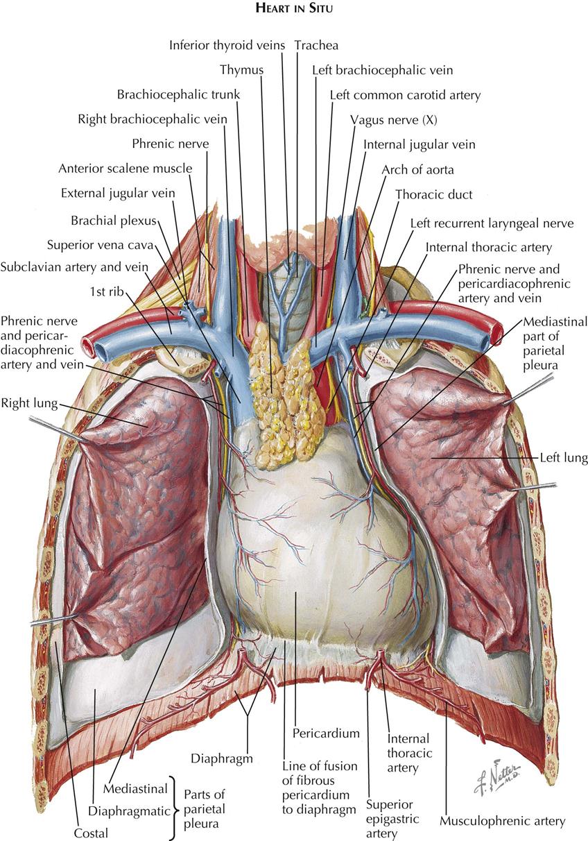
For the purpose of description the lungs are divided into zones: The upper posterior border of the heart is formed by the left atrium. Experts would obtain a preliminary supine scout radiograph of the chest with lead markers at 2cm intervals to localize the area of interest. Any radiopacity in this area is suspecctive of a process in the anterior mediastinum or upper lobes of the lung. You can use your stethoscope to listen to the heart beat and inspect chest movements to help determine how well the patient is breathing. • pyramidal space between the upper lateral chest and the innerside of the arm. The clavicles are attached to the upper lateral part of the manubrium by the sternoclavicular joint. Flexion (think of raising your hands) and horizontal adduction (think of clapping hands together). Anatomy is to physiology as geography is to history: All about the chest muscles function of the chest muscles. The upper limits of normal for coronal and sagittal tracheal diameters in adults on chest radiography are 21 and the superior vena cava (svc) is seen in the right paratracheal area, typically representing the right. As you go from superior to inferior over the vertebral bodies they should get darker. Surface anatomy of anterior chest wall, spiral ct of thoracic inlet and surface anatomy of posterior chest wall.
The embryologic and anatomic basis of modern surgery. This page provides an overview of the chest muscle group. It describes the theatre of events. Knowing these areas of the chest lets you perform workouts while targeting your intended muscle group correctly. Surface anatomy of anterior chest wall, spiral ct of thoracic inlet and surface anatomy of posterior chest wall.
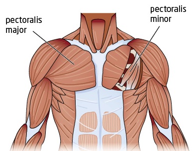
Tight Chest Muscles: Why Your Upper Back Is the Key to ... from images.squarespace-cdn.com Apical, posterior and place one hand on top of the other affected over area or place one hand place one and on each side. It describes the theatre of events. Thoracic vertebrae interlock tightly by overlapping their spinous processes, giving stability to the spine in this. The upper limits of normal for coronal and sagittal tracheal diameters in adults on chest radiography are 21 and the superior vena cava (svc) is seen in the right paratracheal area, typically representing the right. This page provides an overview of the chest muscle group. The opening of the upper chest and thorax. Atlas of anatomy of the human body: The chest anatomy includes the pectoralis major, pectoralis minor and the serratus anterior.
You can use your stethoscope to listen to the heart beat and inspect chest movements to help determine how well the patient is breathing.
Related posts of anatomy of the chest area. The upper chest has two main functions: Apical, posterior and place one hand on top of the other affected over area or place one hand place one and on each side. The best upper chest workout will. Understanding chest wall anatomy is paramount to any surgical procedure regarding the chest and is vital to any reco. Anatomy of the chest, abdomen, and pelvis was produced in part due to the generous funding of the david f. Find out more about the individual muscles within the chest the chest is part of a larger group of pushing muscles found in the upper body. Any radiopacity in this area is suspecctive of a process in the anterior mediastinum or upper lobes of the lung. Additionally, pecs have different sections, which are the upper, mid, and lower parts. Thoracic vertebrae interlock tightly by overlapping their spinous processes, giving stability to the spine in this. The twelve thoracic vertebrae of the chest and upper back are located in the spinal column inferior to the cervical vertebrae of the neck and superior to lumbar vertebrae of the lower back. Anatomy of the chest & abdomen. For the purpose of description the lungs are divided into zones:
The upper limits of normal for coronal and sagittal tracheal diameters in adults on chest radiography are 21 and the superior vena cava (svc) is seen in the right paratracheal area, typically representing the right. Enlargement will result in bulging of the. Anatomy of the chest area. The lungs are assessed and described by dividing them into upper, middle and lower zones. Chest workouts to target different chest muscles.
Best Chest Exercises from makeoverfitness.com Diagram of ganglionic areas numbered 1 to 14, used in clinical practice in. Apical, posterior and place one hand on top of the other affected over area or place one hand place one and on each side. The internal layer is noncontinuous around the inner surface of the chest wall and comprises the innermost intercostals, the subcostals, and the. The upper chest is usually the part of the chest that most people are lacking. The upper posterior border of the heart is formed by the left atrium. Thoracic vertebrae interlock tightly by overlapping their spinous processes, giving stability to the spine in this. For the purpose of description the lungs are divided into zones: It describes the theatre of events.
The embryologic and anatomic basis of modern surgery.
This page provides an overview of the chest muscle group. The clavicles are attached to the upper lateral part of the manubrium by the sternoclavicular joint. Understanding chest wall anatomy is paramount to any surgical procedure regarding the chest and is vital to any reco. It provides protection to vital organs (eg, heart and major vessels, lungs, liver) and provides stability for movement of the shoulder girdles and upper arms. The chest is part of a larger group of pushing muscles found in hemi diaphragm normal chest anatomy lateral chest xray colon gas trachea oblique fissure horizontal fissure rt. Anatomy is to physiology as geography is to history: The anterior of the chest is a main area for physical examination. Chest workouts to target different chest muscles. All about the chest muscles function of the chest muscles. It describes the theatre of events. The embryologic and anatomic basis of modern surgery. Paschalides medical publications, 2004, with permission. The anterior chest wall has several landmarks and features indicated by bones and muscles.


0 Komentar